
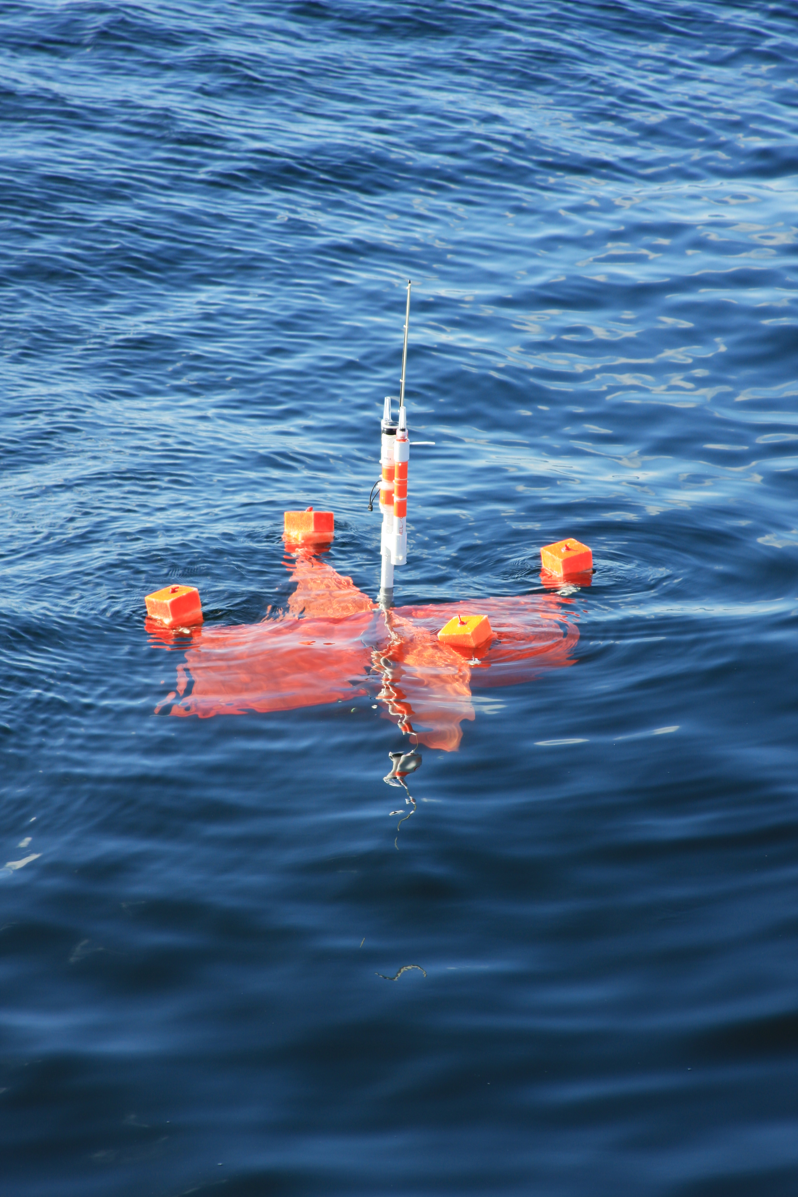
The purpose of this cruise was to examine the dynamics and distribution of the red-tide forming dinoflagellate, Karenia brevis, which forms seasonal blooms on the West Florida Shelf. Led by Cindy Heil (Florida Fish and Wildlife Commission, now at Bigelow Marine Labs), the cruise departed St Petersburg, Florida, and travelled south to the Charlotte River Mouth (near Ft Myers) where a grid patter initiated on a previous cruise leg was completed. The cruise then located a Karenia bloom, placed a drogue in the water, and it was followed for 24 h continuously with regular samplings. Finally, after 6 days we returned to St Petersburg via the Tampa Bay mouth. Ian’s involvement in the cruise focused on three aspects: 1) to examine the presence of Karenia viruses; 2) to conduct induction assays and collect samples for Trichodesmium viruses; and 3) to collect zooplankton for investigations of Nucleopolyhedrovirus diversity.
For Karenia viruses, Ian collected 100 L of water from the highest concentration of Karenia we could find, prepared a virus concentrate which was kept cold for transport back to the labs in St Pete. The virus concentrate was then subject to serial dilution with exponentially growing K. brevis, which was then monitored by Matt Garrett (Florida Fish and Wildlife, and cruise co-chief scientist) for some weeks after the end of the cruise for the abundance of algae. Unfortunately, there was never clearing of K. brevis cultures, even at the lowest virus dilution, suggesting that viral pathogens of the red tide dinoflagellate were not present in the concentrate.
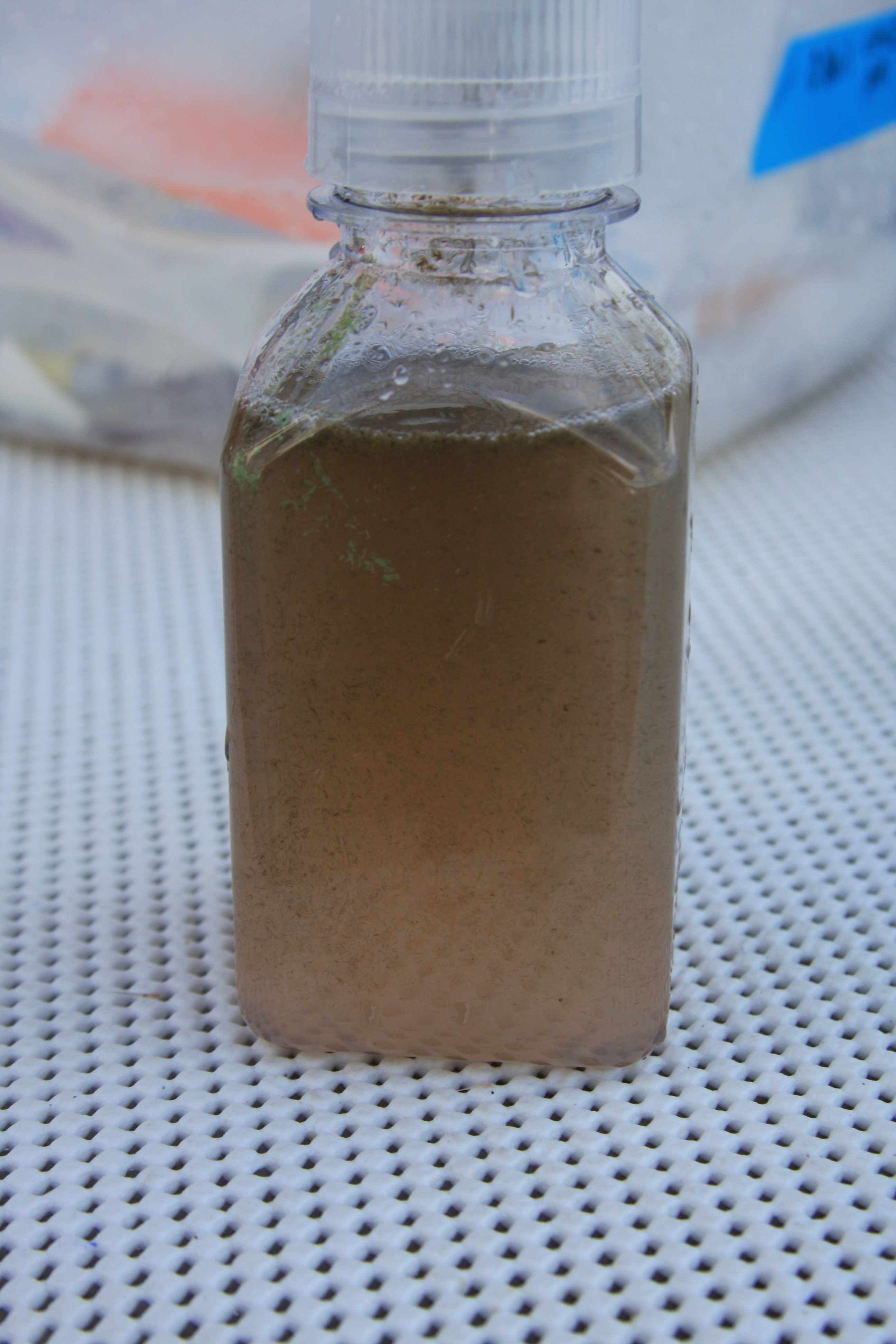
Trichodesmium viruses were investigated by collecting individual trichomes and bulk tows of Trichodesmium from waters during the cruise. These were most dense near the Karenia blooms (T. erythraeum) and at the mouth of the Tampa Bay estuary (T. thiebautii). A goal of this cruise was to collect samples amenable to metaviromic sequencing. To do this, Trichodesmium was collected from several locations; in some cases the mutagen mitomycin C was added to 250ml incubations, while others were kept as controls. After 24 h, the incubations were filtered to remove cell debris and particles and frozen on liquid N2. The filtrate resembled shampoo thanks to the pigment phycoerythrin! Later, back in the lab, the filtrates were prepared for sequencing by Julie Brown (Graduate Student, Microbiology, see separate report).
As part of an exporatory investigation of zooplankton viruses, Ian collected bulk zooplankton and individual animals from several locations. The plankton in many cases was dominated by pteropods, however there were also abundant calanoid copepods, and in the Trichodesmium blooms there was Macrosetella present. These were frozen and transported to the lab, where they were analyzed by PCR for the presence of Nucelopolyhedroviruses, a group of insect viruses that form environmental degradation-resistant polyhedral, that we hypothesized would be present in zooplankton. However, our results ultimately showed that these were not present in the zooplankton, at least ones we could pick up by our primer sets.


Publications from this Expedition:
Brown JM, LaBarre BA, Hewson I (2013) “Characterization of Trichodesmium-associated viral communities in the eastern Gulf of Mexico” FEMS Microbiology Ecology 84: 603-613
Eaglesham JB, Hewson I (2013) “Widespread detection of eukaryotic circular ssDNA viral genotypes in estuarine, coastal and open ocean net plankton” Marine Ecology Progress Series 494: 55-64
