
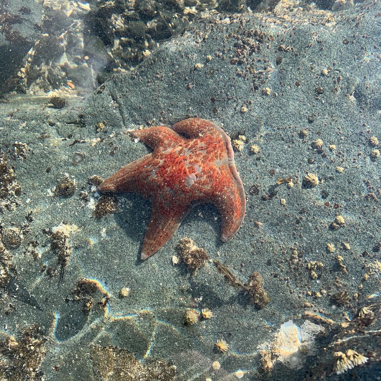
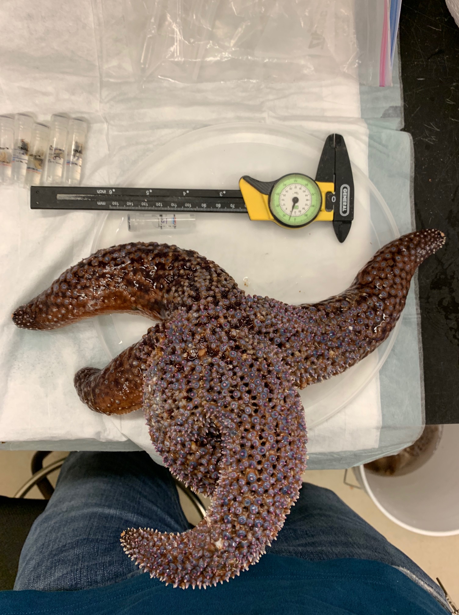
The results of work in 2018 and 2019 suggested a key role of microorganisms inhabiting the sea star-seawater interface. But several important questions remained. Most importantly: if this effect occurs on all sea stars, why were some species more impacted than others (and some species not affected at all?). Boundary layers, which occur on all aquatic surfaces, are established by water flow and turbulence, and are a function of the height of surface features and their corrugatedness (known as rugosity). We hypothesized that inter-species variation in SSW susceptibility may relate to their relative rugosity – some species, like bat stars (Patiria miniata) and leather stars (Dermasterias imbricata) experienced few reports of SSW – these species had relatively smooth dorsal surfaces compared to more heavily affected species, like Pisaster ochraceus and Pycnopodia helianthoides. Quantifying rugosity, however, is a challenge. In consultation with Ian Porter at Cornell’s College of Veterinary Medicine, we designed a survey to compare the relative rugosity (and their surface area:volume) by computed tomography, which is normally reserved for clinical specimens like farm and domestic animals. However, we needed intact, preserved specimens with which to work!
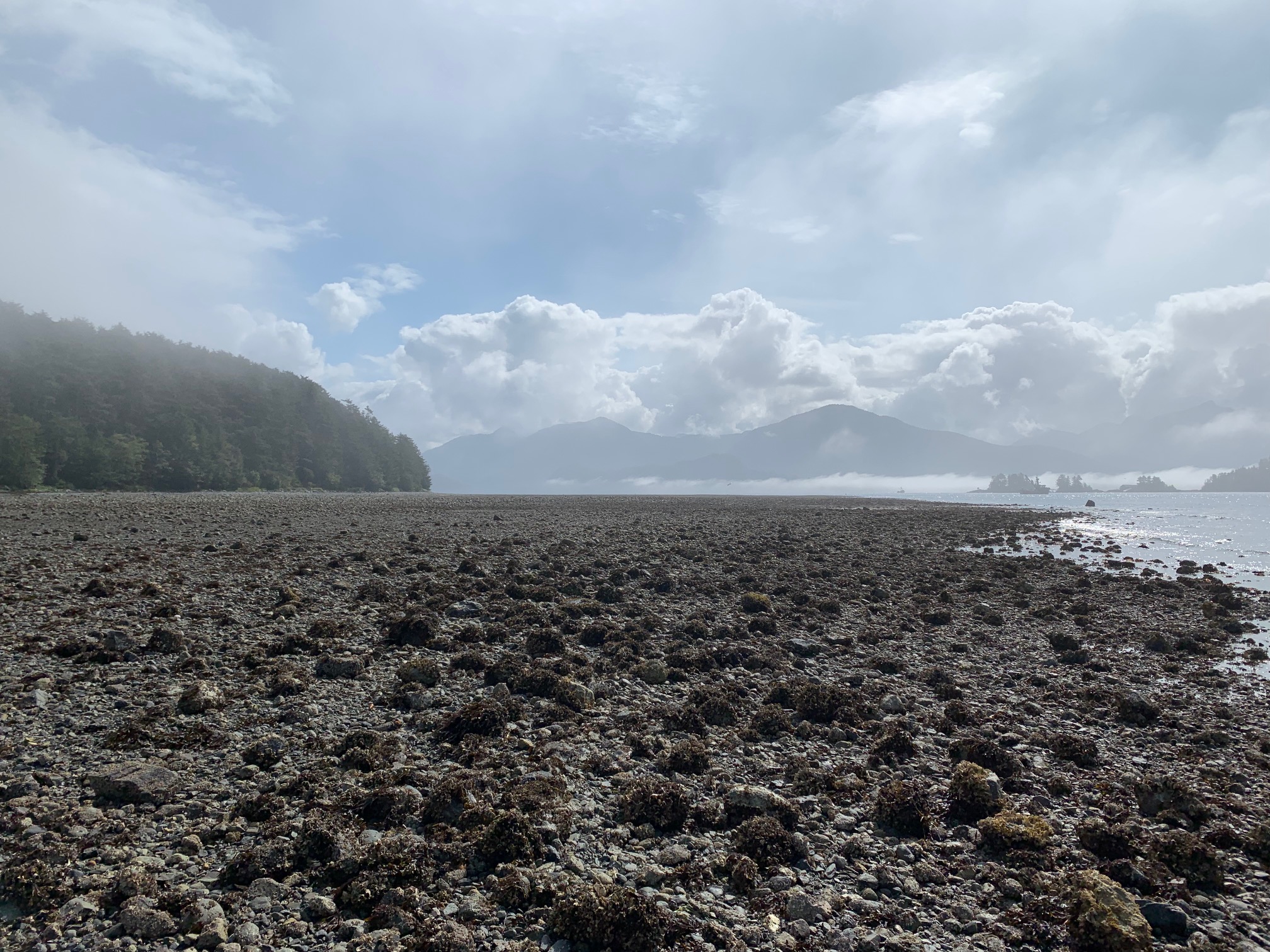
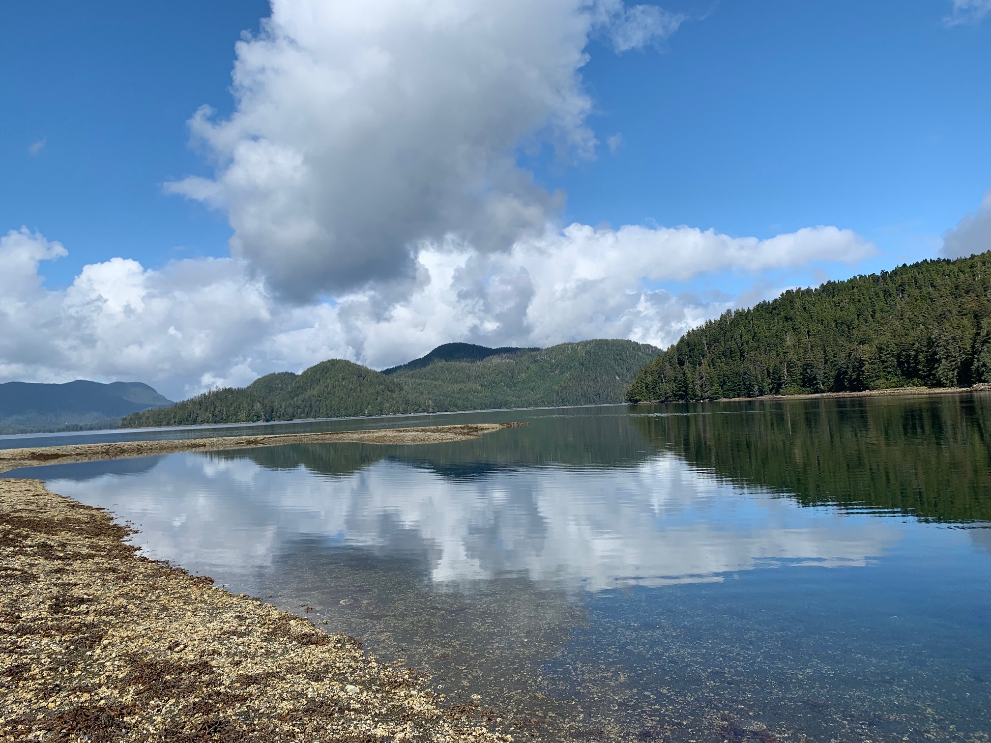
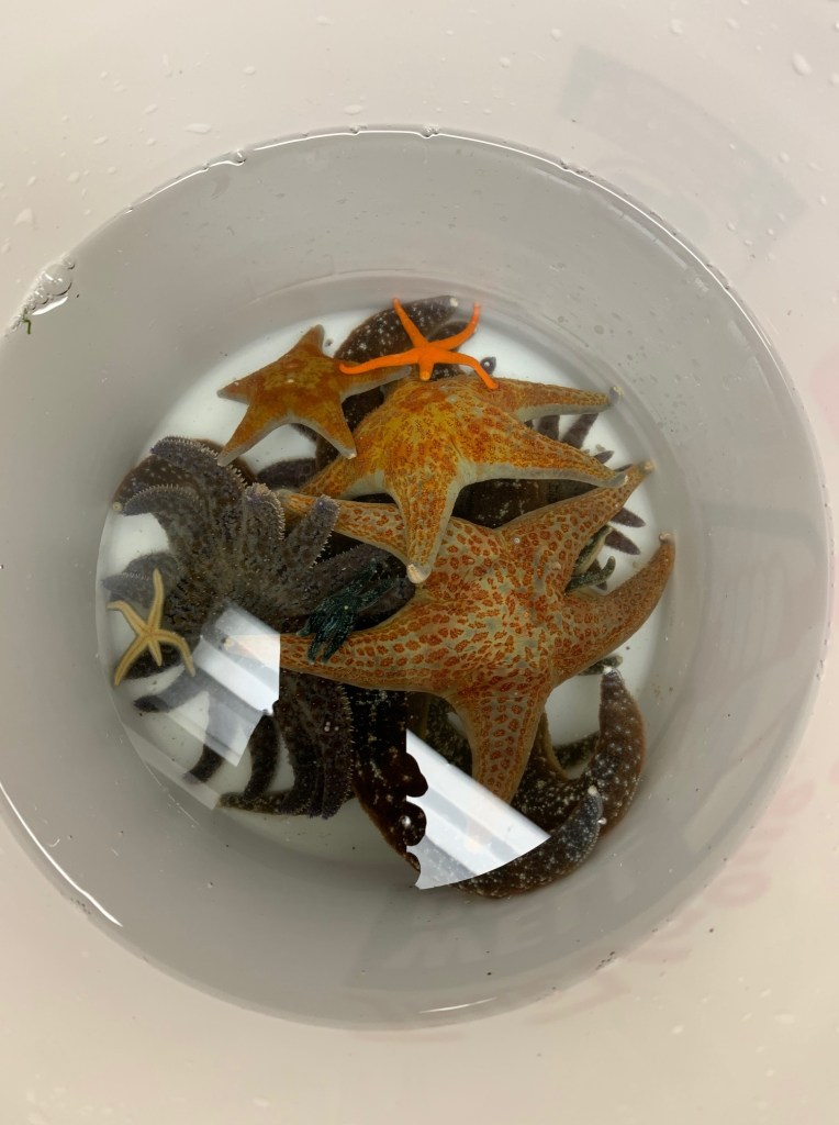
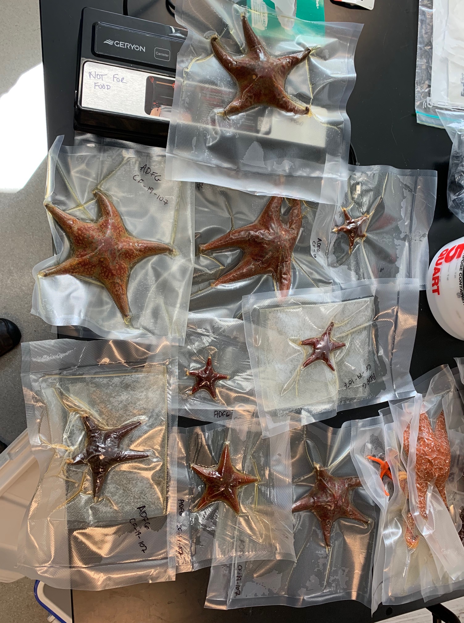
In September 2019, Ian traveled to Sitka, Alaska, to collect intact specimens for this work. First, he collected several specimens by hand within the intertidal zone, and transported them to the University of Alaska Southeast, where he was kindly hosted by Joel Markis and Marnie Chapman. Taylor White, a graduate student at UC Santa Cruz, also collected specimens subtidally at a couple of offshore sites by SCUBA. In the lab, specimens were fixed, and prepared for shipment to Cornell. One of the most beautiful sampling sites we’ve ever sampled!
Later that month, Ian traveled to Santa Cruz to process specimens of several southern species (Pisaster giganteus, Leptasterias hexactis and Patiria miniata) that had been collected by Pete Raimondi’s group. In addition, specimens were collected by Joe Gaydos (UC Davis) at the Friday Harbor Marine Labs.
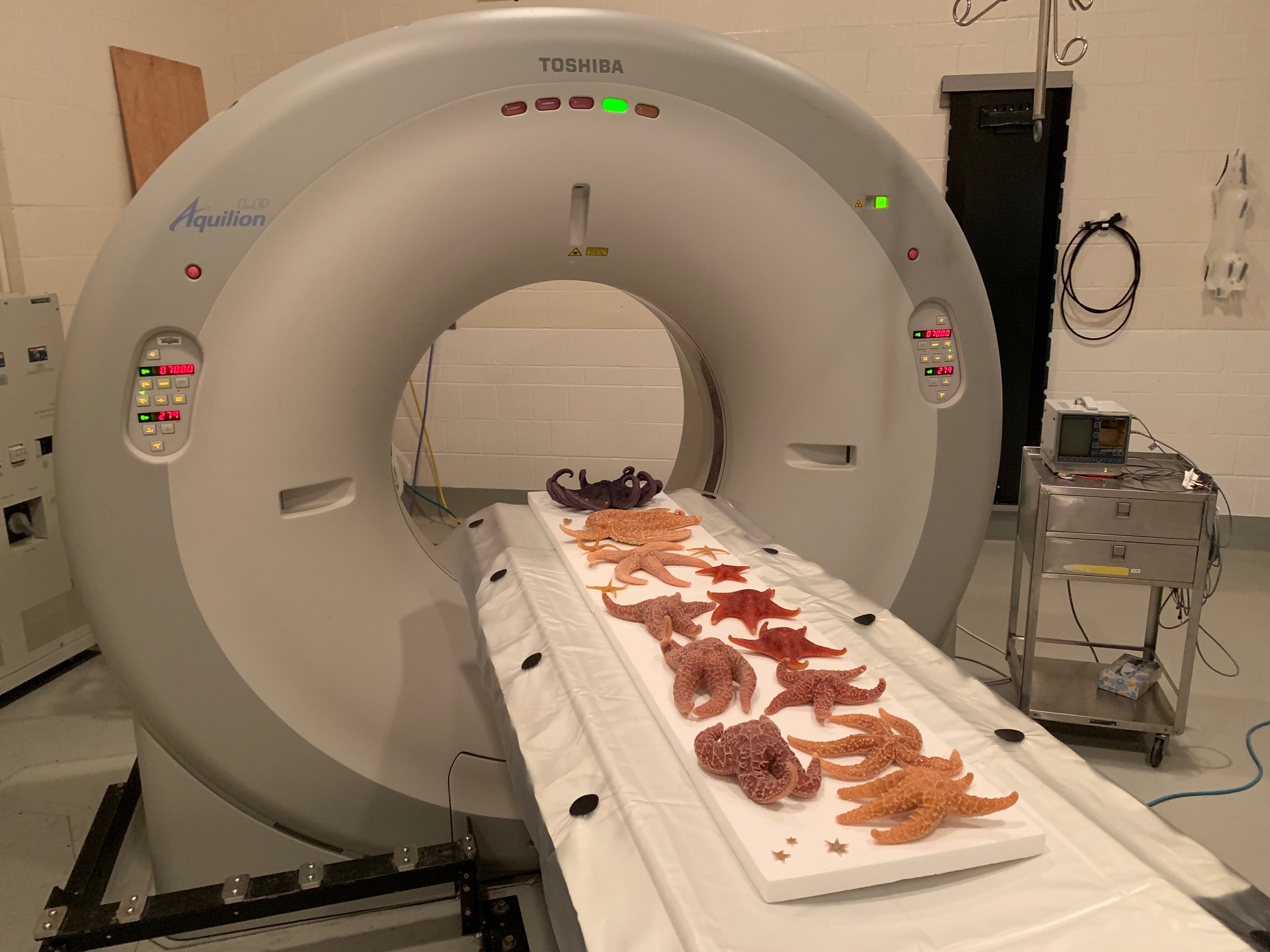
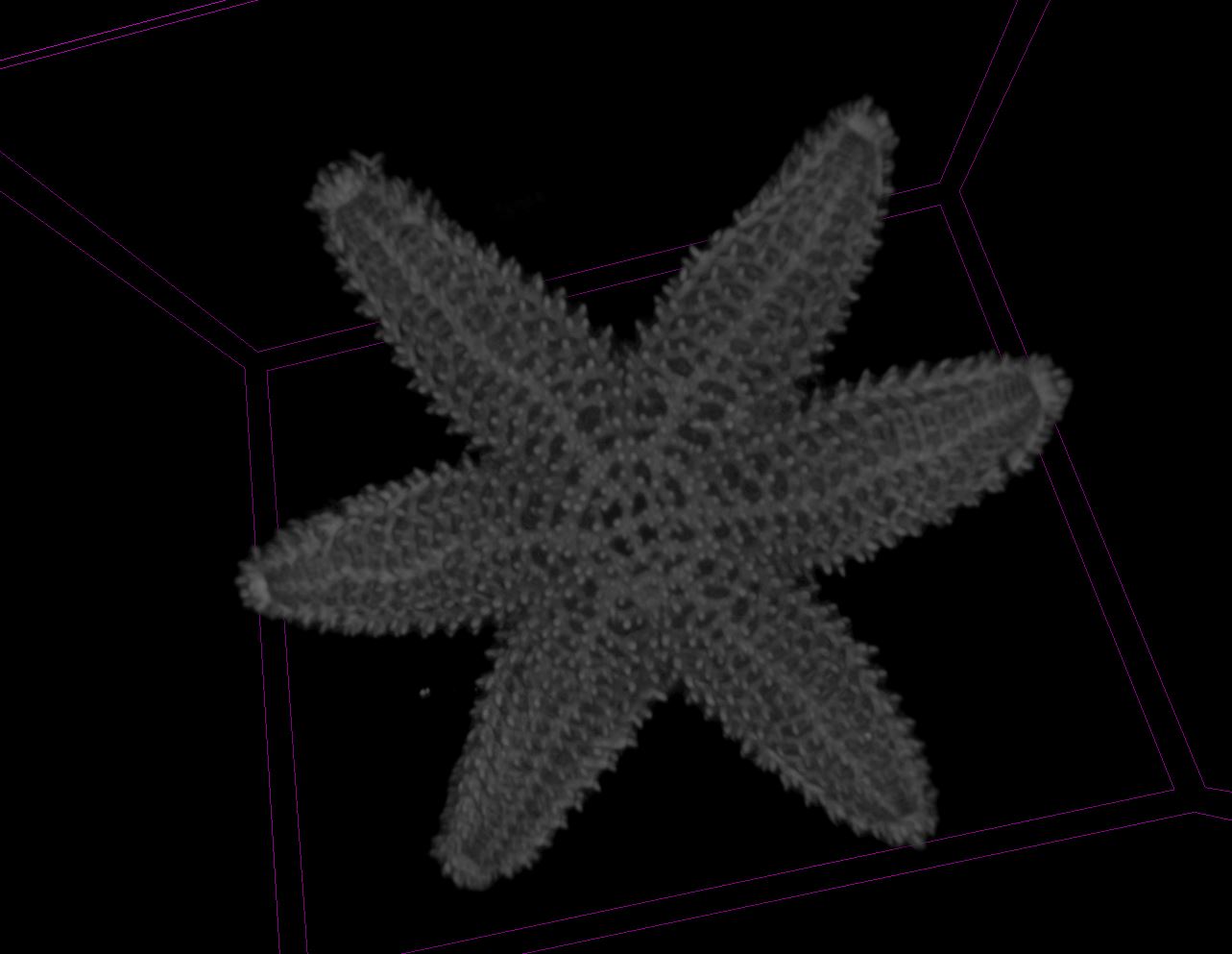
Once back in Ithaca, the specimens were subject to large animal CT scans, which provided important information about their physical features. However, we soon noticed that many surface features of specimens were larger than the resolution of the large animal CT, and so we turned to the finer resolution micro-CT at Cornell’s imaging facility (Cornell Biotechnology Resource Center).
The results of this work demonstrated that more heavily SSW-affected species were more rugose than less heavily affected or not affected species. Details of this work can be found here.
Publications from this Expedition:
Aquino CA, Besemer RM, DeRito CM, Kocian J, Porter IR, Raimondi P, Rede JE, Schiebelhut LM, Sparks JP, Wares JP, Hewson I (2021) “Evidence that microorganisms at the animal-water interface drive sea star wasting disease” Frontiers in Microbiology. DOI: https://www.frontiersin.org/articles/10.3389/fmicb.2020.610009/
